Spina bifida is a neural tube defect that occurs when the spine and spinal cord fail to close properly during the first month of pregnancy. Detecting it before birth gives doctors and families a chance to plan treatment, reduce complications, and make informed choices.
Key Points
- Spina bifida can be spotted as early as 11‑13 weeks with a detailed ultrasound.
- Maternal serum alpha‑fetoprotein (AFP) is a reliable blood marker for neural tube defects.
- Fetal MRI adds clarity when ultrasound findings are ambiguous.
- Folic acid intake before conception and during early pregnancy cuts the risk by up to 70%.
- Early detection opens doors to prenatal surgery and tailored postnatal care.
Understanding Spina Bifida
Spina bifida belongs to a broader group called neural tube defects (birth defects of the brain, spine, or spinal cord caused by incomplete closure of the embryonic neural tube). The condition ranges from mild (occult spina bifida) to severe forms like myelomeningocele, where the spinal canal remains open, exposing nerves and spinal fluid.
When the defect is identified early, a multidisciplinary team-obstetricians, pediatric neurosurgeons, genetic counselors-can map out a care plan that targets the specific level of the spine involved, the presence of associated brain anomalies, and the expected functional outcomes.
Why Early Detection is Critical
Finding spina bifida in the first trimester changes the entire trajectory of pregnancy management. First, it gives parents time to seek credible information and emotional support before the pregnancy progresses. Second, it allows doctors to schedule targeted follow‑up imaging (like fetal MRI at 20‑24 weeks) that refines the diagnosis.
Most importantly, early detection enables the option of prenatal surgery. Studies from the Management of Myelomeningocele Study (MOMS) show that repairing the defect before 26 weeks reduces the need for shunt placement and improves motor outcomes in the first year of life. Without early diagnosis, families lose that window.
Screening Tools and When They’re Used
Multiple tests complement each other to catch spina bifida with high confidence.
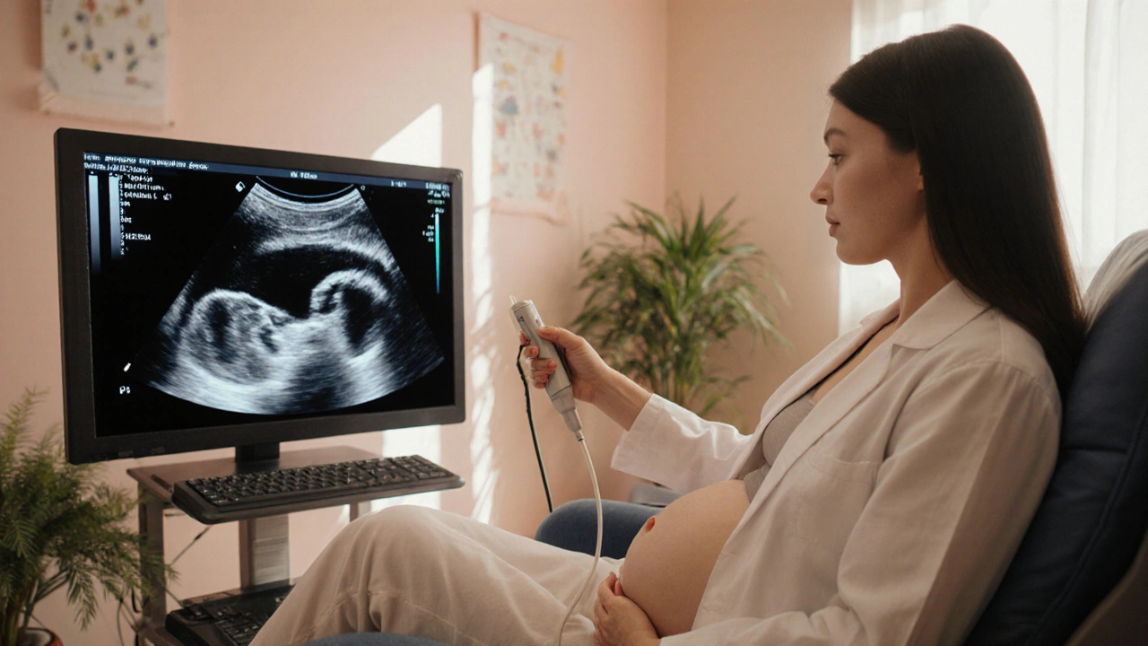
- Ultrasound (high‑frequency sound waves that produce real‑time images of the fetus) is the frontline tool. A nuchal translucency scan at 11‑13 weeks can spot the “banana sign” or elevated posterior fossa that hint at open spina bifida. A detailed anatomy scan at 18‑22 weeks confirms the defect and assesses spinal level.
- Maternal serum alpha‑fetoprotein (AFP) (a protein produced by the fetal liver; high levels in maternal blood suggest open neural tube defects) is drawn between 15‑20 weeks. Values above the 2.5‑multiple‑of‑the‑median (MoM) threshold raise red flags and trigger follow‑up imaging.
- Fetal MRI (magnetic resonance imaging that provides detailed soft‑tissue contrast without ionizing radiation) is reserved for ambiguous ultrasound cases or when surgeons need precise anatomy for planning prenatal repair.
- Amniocentesis (a needle‑based sampling of amniotic fluid to test for genetic conditions and AFP levels) is invasive but can confirm AFP elevations and rule out chromosomal abnormalities that often accompany severe neural tube defects.
| Method | Optimal Timing | Detection Rate | Invasiveness |
|---|---|---|---|
| Obstetric Ultrasound | 11‑13 wk (nuchal), 18‑22 wk (anatomy) | ≈85‑90% | Non‑invasive |
| Maternal Serum AFP | 15‑20 wk | ≈70‑80% | Non‑invasive (blood draw) |
| Fetal MRI | 20‑24 wk | ≈95% (when needed) | Non‑invasive |
| Amniocentesis | 15‑18 wk | Confirmatory | Invasive (≈0.5% miscarriage risk) |
Risk Factors and Prevention
The single most effective preventive measure is folic acid (a B‑vitamin (vitamin B9) essential for DNA synthesis and neural tube closure). The CDC recommends 400µg daily for all women of childbearing age, and 600µg for those actively trying to conceive. Randomized trials show a 50‑70% reduction in spina bifida incidence with proper supplementation.
Additional risk contributors include:
- Maternal diabetes (poorly controlled blood glucose)
- Obesity (BMI>30)
- Use of certain anti‑seizure medications (e.g., valproic acid)
- Family history of neural tube defects
Addressing these factors early-optimizing glycemic control, achieving a healthy weight, discussing medication alternatives with a physician-lowers the odds of the defect.

Management Options After Diagnosis
Once spina bifida is confirmed, the care pathway splits into three main branches:
- Prenatal surgery: Performed between 19‑26 weeks via open fetal repair or minimally invasive fetoscopic techniques. Candidates typically have isolated myelomeningocele without severe associated brain anomalies.
- Expectant management: Some families opt to continue the pregnancy without fetal intervention, focusing on postnatal surgery and rehabilitation.
- Termination: In jurisdictions where legal and ethically permissible, severe forms with poor neurological prognosis may lead families to consider this option.
All three routes require thorough counseling. A pediatric neurosurgeon evaluates the defect’s level (e.g., L1‑L2 versus L4‑L5) because that predicts motor function and bladder control. A neonatologist reviews potential complications like hydrocephalus, which affects about 80% of myelomeningocele cases.
Decision‑Making and Counseling
Genetic counseling provides clarity on recurrence risk-approximately 2-5% for a second child if the first was affected, rising to 10% with a family history. Counselors also explain the long‑term needs: physical therapy, orthopedic interventions, and sometimes lifelong bladder management.
Emotional support is just as vital. Connecting with parent groups, such as the Spina Bifida Association, can reduce anxiety and provide real‑world expectations.
Practical Checklist for Expectant Parents
- Start a daily prenatal vitamin with at least 400µg folic acid before conception.
- Schedule a first‑trimester ultrasound (11‑13 weeks) and discuss the possibility of an early anatomy scan.
- If AFP results are elevated, arrange a follow‑up detailed ultrasound and consider fetal MRI.
- Meet with a multidisciplinary team (maternal‑fetal medicine, neurosurgery, genetics) as soon as the defect is identified.
- Ask about eligibility for prenatal repair-timing, surgical center location, and insurance coverage.
- Plan for postnatal care: identify a neonatal intensive care unit with neurosurgical capability.
Next Steps After Reading This
If you’re already pregnant and worried about spina bifida, contact your obstetrician to schedule the recommended screening timeline. If you’re planning a family, start folic‑acid supplementation today and discuss any existing health conditions that could raise risk.
Early detection isn’t just a medical checkbox; it reshapes outcomes, reduces complications, and gives families a clearer roadmap for the months ahead.
Frequently Asked Questions
Can spina bifida be cured after birth?
Surgical closure of the defect is standard soon after birth, but the term “cure” is misleading. Surgery prevents infection and further nerve damage, yet many children still need lifelong therapies for mobility, bladder control, and possible hydrocephalus management.
How accurate is the AFP blood test?
AFP is a sensitive screening tool but not definitive. Elevated levels flag a potential open neural tube defect in about 70‑80% of cases, prompting imaging for confirmation. False positives can occur due to dating errors, multiple pregnancies, or maternal liver disease.
Is fetal MRI safe for the baby?
Yes. Fetal MRI uses magnetic fields, not ionizing radiation, and has been shown to be safe when performed after 20 weeks gestation. The main limitation is the need for the mother to stay still for the duration of the scan.
What are the long‑term outcomes for children who had prenatal surgery?
Data from the MOMS trial indicates better motor function at 30 months, fewer shunt procedures for hydrocephalus, and improved ambulation rates compared with postnatal repair. However, risks include preterm birth and uterine scarring for the mother.
Should I take higher doses of folic acid if I have a family history of spina bifida?
Many experts recommend 4mg (4000µg) daily for women with a previous child affected by a neural tube defect or a known hereditary risk. Discuss the dosage with your healthcare provider to avoid masking vitamin B12 deficiency.
Early detection of spina bifida can mean the difference between a life‑changing surgery and a manageable condition. Stay proactive, ask questions, and lean on specialized teams-you deserve the best possible outcome for both you and your baby.


Marry coral
September 28, 2025 AT 06:52Spina bifida? Yeah, that’s a huge red flag. Get that ultrasound ASAP, don’t wait for the doctor to be lazy. Early surgery can actually save a lot of trouble later.
Emer Kirk
October 7, 2025 AT 22:24Honestly feels like the world forgets how scary this can be for moms, the waiting, the uncertainty, the whole nightmare.
Roberta Saettone
October 17, 2025 AT 13:56Great rundown, but let’s be real – most folks still ignore the folic acid tip until after they’re already pregnant. If you’re not popping those pills now, you’re basically gambling with your baby’s spine. Also, that “banana sign” thing? Cute nickname, not a miracle cure.
Sue Berrymore
October 27, 2025 AT 04:28Listen, you’re not alone in this roller‑coaster! Grab a support group, talk to a genetic counselor, and remember that early detection is the secret weapon that can turn a scary diagnosis into a manageable plan. Every step you take now builds a stronger foundation for the little one’s future!
Jeffrey Lee
November 5, 2025 AT 19:59Look folks the data is crystal clear – folic acid works and the US should push it harder than any other country stop doddling around the facts.
Ian Parkin
November 15, 2025 AT 11:31It is most commendable that you have taken the initiative to understand such a complex condition; nevertheless, one must consider the broader public health implications-particularly the necessity for universal fortification programs. Apologies for any typographical oversight.
RONEY AHAMED
November 25, 2025 AT 03:03Sounds like a solid plan, just make sure you stick to the screening schedule.
emma but call me ulfi
December 4, 2025 AT 18:35I think staying calm and following the doctor’s timeline is probably the best way to handle everything.
George Gritzalas
December 14, 2025 AT 10:07Wow, another checklist! Because who doesn’t love a bullet‑point list at 2 AM, right? Let’s hope the paperwork doesn’t outweigh the actual care.
Alyssa Matarum
December 24, 2025 AT 01:38Key takeaway: early AFP + ultrasound = earlier options.
Lydia Conier
January 2, 2026 AT 17:10First off, congratulations on doing the homework – spina bifida is not something you just stumble onto without a reason.
Early detection is like having a roadmap before you set out on a treacherous hike.
When doctors spot the defect at 11‑13 weeks they can schedule the necessary follow‑ups before the pregnancy gets too far along.
This means you can meet with a maternal‑fetal medicine specialist, a neurosurgeon, and a genetic counselor all in one coordinated plan.
Having the whole team on board early often cuts down on the anxiety that builds up when you’re left in the dark for months.
The MOMS trial showed that prenatal repair before 26 weeks slashes the need for shunt placement later on, which is a huge quality‑of‑life win.
Sure, the surgery itself carries risks like preterm birth, but those are weighed against the potential lifelong mobility benefits.
Don’t forget the power of folic acid – a simple daily pill can drop the risk by up to 70 %, which is something no one should overlook.
If you have a family history, talk to your doc about a higher dose, like 4 mg, because the standard 400 µg might not be enough.
Also, managing diabetes, maintaining a healthy weight, and reviewing any anti‑seizure meds can make a big differecne.
When the ultrasound shows the “banana sign” or the “lemon sign” you’re already ahead of the curve.
If the picture is unclear, a fetal MRI can give crystal‑clear images without radiation.
All this info can feel overwhelming, so write down questions, bring a friend to appointments, and use reputable online forums for extra support.
Remember that every pregnancy is unique, so the final plan will be tailored to your baby’s exact spinal level and any associated brain findings.
Bottom line: early detection turns a scary unknown into a series of actionable steps, and that empowerment can make all the difference for you and your little one.
ruth purizaca
January 12, 2026 AT 08:42Such a perfunctory overview hardly scratches the surface of clinical nuance.
Shelley Beneteau
January 22, 2026 AT 00:14I’m curious how different healthcare systems worldwide handle prenatal screening for spina bifida, especially in places where folic‑acid fortification isn’t mandatory.
Sonya Postnikova
January 31, 2026 AT 15:45You’ve got this! 🌟 Keep pushing for those early appointments and surround yourself with a solid team 😊
Anna Zawierucha
February 10, 2026 AT 07:17Wow, a checklist that reads like a novel – because who doesn’t love turning prenatal care into a literary masterpiece?
Mary Akerstrom
February 19, 2026 AT 22:49I hear you, it’s a lot to take in and you’re doing the best you can
Delilah Allen
March 1, 2026 AT 14:21Consider this: every decision you make now is a thread in the tapestry of your child’s future; neglecting early detection is tantamount to severing those threads before they even begin; therefore, act decisively!
Nancy Lee Bush
March 11, 2026 AT 05:52Could early detection also open doors to cutting‑edge research trials that might benefit your baby? The possibilities are exciting; let’s stay hopeful and keep exploring! 😊